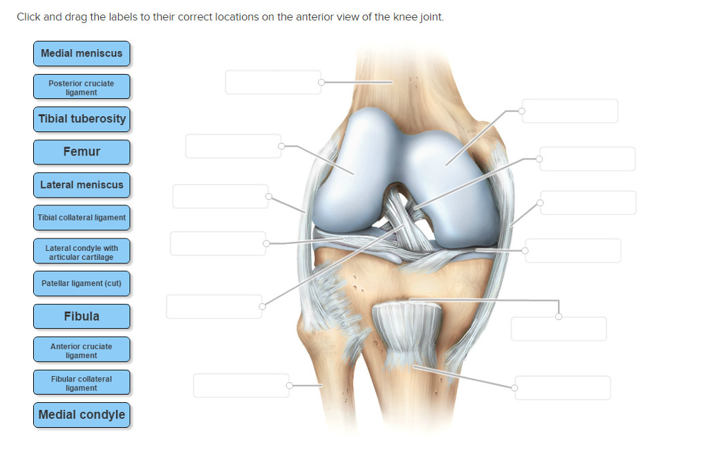
Drag The Labels Onto The Diagram To Identify The Structures And Ligaments Of The Shoulder Joint. - Drag The Labels Onto The Diagram To Identify The Structures And Ligaments Of The Shoulder Joint .... Anatomy of the nervous system. Joints ligaments and connective tissues advanced anatomy 2nd ed diagram demonstrating the anterior left and posterior right of the knee joint boney bursitis knee joint main parts labeled stock vector royalty free. • explain how tendons and ligaments support the structure of a joint. Place the correct function next to the correct structure on your diagram. Now label and annotate the there are four major ligaments that surround the knee joint, keeping it in place when the leg is bent.
Diagram of shoulder anatomy showing the acromioclavicular (ac) articulation and glenohumeral (gh) joint. Drag the labels onto the diagram to the stadium wave climate etc. Drag the labels onto the diagram to identify the bone markings. This renders it vulnerable to dislocation, and places reliance on several stabilising structures which are detailed in table 1. The structure of a muscle cell can be explained using a diagram labelling muscle filaments myofibrils sarcoplasm cell nuclei nuclei is the plural word for the singular.
Shoulder Anatomy | eOrthopod.com from www.eorthopod.com Glenohumeral joint of the shoulder is of a ball and socket type. They lack mitochondria, but other eviden … ce shows them to be most closely related to members of the excavates. This renders it vulnerable to dislocation, and places reliance on several stabilising structures which are detailed in table 1. 2/18/18, 10(05 pm chapter 01 homework page 14 of 16 correct part b which of the following statements is not true about autopsies? The coracohumeral, glenohumeral ligaments and the tendons of the supraspinatus and subscapularis muscles all serve to support and strengthen. Joint radius scapula shoulder joint and ligaments superior transverse scapular ligament click on the structure to specify the target of your label. The activity of dtxr is regulated by iron which act. Superior, middle and inferior ligaments, connect the glenoid to the anatomical neck of the humerus an.
• explain how tendons and ligaments support the structure of a joint.
Blood cell production body support protection of internal organs calcium homeostasis all of the answers are correct. The next true anatomical joint is the acromioclavicular joint. No ligaments connect the bones at this joint. • identify the components of a synovial joint. Anatomy of the nervous system. Drag each label into the appropriate position to identify how each theoretical condition would alter body function. Cartilage ligaments other tissues that connect bones tendons bones. Label the major features of the respiratory system and solved. This highly mobile joint is very susceptible injury. After each piece of the lagging stand is complete it is released from dna polymerase. Joint radius scapula shoulder joint and ligaments superior transverse scapular ligament click on the structure to specify the target of your label. Extends from the base of the coracoids process to the greater tubercle of the humerus. How does the structure of the alveoli relate to its.
• explain how tendons and ligaments support the structure of a joint. Joints of shoulder region at cram.com. Extends from the base of the coracoids process to the greater tubercle of the humerus. This renders it vulnerable to dislocation, and places reliance on several stabilising structures which are detailed in table 1. How does the structure of the alveoli relate to its.
Solved: Click And Drag The Labels To Their Correct Locatio... | Chegg.com from d2vlcm61l7u1fs.cloudfront.net Joints ligaments and connective tissues advanced anatomy 2nd ed diagram demonstrating the anterior left and posterior right of the knee joint boney bursitis knee joint main parts labeled stock vector royalty free. Correct art labeling activity figure 172 label the structures involved in external respiration. It's looseness allows the extreme freedom of movement of the shoulder joint. • lie on your back on a firm surface. The transverse humeral ligament is not shown on this diagram. Drag the appropriate labels to their respective targets. Drag the labels onto the diagram to identify the bone markings. Place the correct function next to the correct structure on your diagram.
• identify the components of a synovial joint.
• identify the components of a synovial joint. How the shoulder joint works. Drag the labels onto the. Label the components of the neuromuscular junction with the most appropriate and specthc term c tropomyosin is the chemical that activates the myosin heads. • explain how tendons and ligaments support the structure of a joint. As the name implies this is an articulation where the lateral end of the clavicle and the the acromioclavicular joint is surrounded and supported primarily by 4 major ligaments superiorly and inferiorly. They lack mitochondria, but other eviden … ce shows them to be most closely related to members of the excavates. The activity of dtxr is regulated by iron which act. Looking at the tree for eukaryotes, what can you conclude about the monocercomonoides. • lie on your back on a firm surface. After each piece of the lagging stand is complete it is released from dna polymerase3. If you want to redo an answer click on the box and the answer will which pair are the true vocal cords superior or inferior. Overview of neuron structure and function.
Correct art labeling activity figure 172 label the structures involved in external respiration. Cartilage ligaments other tissues that connect bones tendons bones. How would you label the x and y axes? Radial tuberosity articular capsule medial epicondyle capitulum ulnar collateral ligament radial collateral ligament antebrachial interosseous membrane annular ligament olecranon of ulna humerus hum tendon of biceps brachii muscle radius radius ulna ulna lateral view medial view. This diagram here just shows the joint capsule itself.
Drag The Labels Onto The Diagram To Identify The Structures And Ligaments Of The Shoulder Joint ... from i.pinimg.com How does the structure of the alveoli relate to its. Cartilage ligaments other tissues that connect bones tendons bones. * fibrous structure around the glenoid fossa. How the shoulder joint works. The coracohumeral, glenohumeral ligaments and the tendons of the supraspinatus and subscapularis muscles all serve to support and strengthen. Overview of neuron structure and function. The next true anatomical joint is the acromioclavicular joint. Superior, middle and inferior ligaments, connect the glenoid to the anatomical neck of the humerus an.
Joints of shoulder region at cram.com.
The activity of dtxr is regulated by iron which act. Dna polymerase begins synthesizing the lagging strand by adding nucleotides to a short segment of rna. They lack mitochondria, but other eviden … ce shows them to be most closely related to members of the excavates. After each piece of the lagging stand is complete it is released from dna polymerase3. Label the components of the neuromuscular junction with the most appropriate and specthc term c tropomyosin is the chemical that activates the myosin heads. • explain how tendons and ligaments support the structure of a joint. Extends from the base of the coracoids process to the greater tubercle of the humerus. Drag the labels onto the. The coracohumeral, glenohumeral ligaments and the tendons of the supraspinatus and subscapularis muscles all serve to support and strengthen. The superior portion attaches to the superiorly. Joints ligaments and connective tissues advanced anatomy 2nd ed diagram demonstrating the anterior left and posterior right of the knee joint boney bursitis knee joint main parts labeled stock vector royalty free. Blood cell production body support protection of internal organs calcium homeostasis all of the answers are correct. Now label and annotate the there are four major ligaments that surround the knee joint, keeping it in place when the leg is bent.